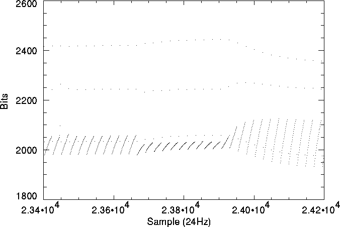
Figure 5.3: Example of memory effects
The band 2 and 4 detectors used in SWS ``remember'' their previous illumination history - bands 1 and 3 are not affected by memory effects. Going from low illumination to high illumination, or vice-versa, results in the detectors asymptotically reaching a value. These are referred to as ``memory effects''. Currently the only memory effects accounted for in the pipeline are those that affect dark currents - the first few dark current readouts are ignored.
An example of memory effects can be seen in Figure 5.3, which shows ERD data from detector 39 (band 4) as the illumination changes during a dark current check. It can be seen that the detector takes a few resets to stabilise both on light-dark or dark-light illumination changes.

Figure 5.3: Example of memory effects
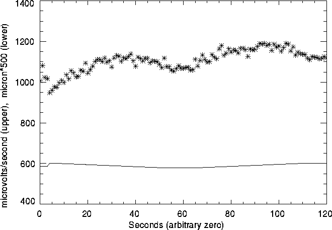
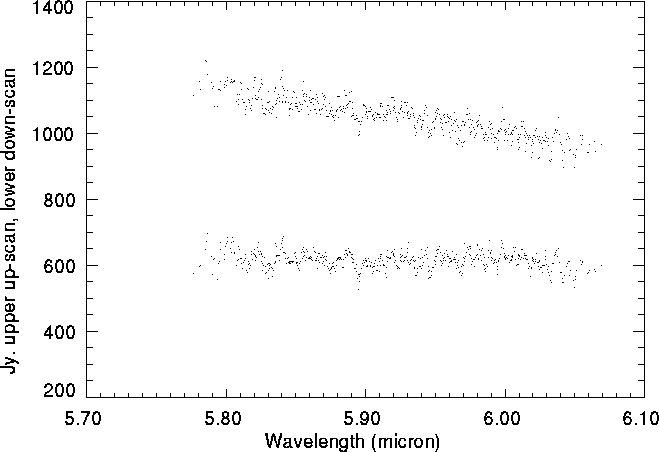
Another example of memory effects is the effect seen in up-down scans of fairly bright sources. For sources with fluxes greater than about 1000 Jy memory effects cause the up and down scans to differ in response (and hence flux) by up to 20% in bands 2 and 4. This effect can be seen in figures 5.4 and 5.5. Figure 5.4 shows the output of detector 13 against time along with the wavelength of light falling on the detector. It can be seen that the detector output rises from a low state at the start of the observation, and that it does not fall back to its low level at the end. Figure 5.5 shows the result of this in AAR. The top plot is for the up-scan, the lower for the down-scan - the down scan is shifted down by 500 Jy for clarity and remember that the up-scan actually scans down in wavelength (see section 8.3.6). While the short wavelength data for both scans has approximately the same flux level, the long wavelength data (that taken at the start and end of the observation) has quite different flux levels. Realistically, only the down scan should be used.
It is important to stress this - if you see your data is affected you must decide how much data to use and in extreme cases use only one scan.
Memory effects can also change the shape of line profiles. As an example, as the grating scans across a strong gaussian line the sensitivity of the detectors will change. If this change is large enough it will result in one side of the gaussian having a detectably different shape from the other. As lines are scanned in different directions by the up-down scans , the line will have a different shape in both scans. If the scans are averaged the line will be broader than it actually is. Users should check for these effects and if they occur only use the unaffected half of a scan. These effects mainly happen in band 4, due to it's larger memory effects.

Figure 5.4: Example of memory effects on up-down scans as seen in
SPD. Top is the detector output, bottom is the wavelength seen by that
detector.

Figure 5.5: Example of memory effects on up-down scans as seen in
AAR. The down scan has been shifted down by 500 Jy for clarity.
Table 5.2 (based on ground based tests) gives indications of the errors introduced into flux calibration by memory effects.
| 1 InSb | 2 Si:Ga | 3 Si:As | 4 Ge:Be | 5 Si:Sb | 6 Ge:Be |
| | | | | | |